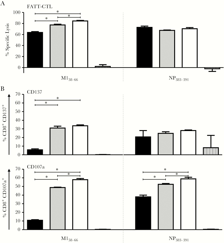
Figure 2.
Activation and lytic activity of M158-66- and NP383-391-specific CD8+ T lymphocytes (CTLs) after stimulation with matrix protein 1-nucleoprotein-enhanced green fluorescent protein (M1-NP-eGFP) transfected cells. M158-66- and NP383-391-specific CTLs were incubated with HLA-matched target cells transfected with chimeric M1-NP-eGFP fusion plasmids that encode the M1 protein of H3N2s1994 (black), H1N1pdm09 (gray) or H1N1pdm09/2016 (white) influenza A viruses, and lytic activity was determined by fluorescent antigen-transfected target cell (FATT)–CTL assay (A). In addition, expression of activation markers CD137 and CD107a by the respective CTLs was assessed (B). GFP (A) or mock (B) transfected stimulator cells served as a negative control (hatched bars). Data points represent the mean, and error bars indicate the standard deviation of quadruplicates (n = 4). * Indicates statistically significant differences between groups after correction for multiple hypothesis testing using a false discovery rate of 0.01.

