
Fig. 5: Electrical activity as a neuronal function of the BMPS.
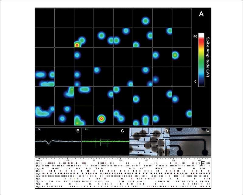
Cells were cultured in 3D for 8 weeks and then cultured in 48-well MEA plates for 2 more weeks. (A) Heat map recordings from a 48-well plate. Illustration of an active well showing (B) spike morphology and (C) spike activity. (D and E) Phase-contrast imaging of the BMPS on MEAs, electrode diameter is 40–50 μm and inter-electrode space is 350 μm. (F) Activity pattern recordings over 0.05 spikes/sec of the electrode over 10 min.