Figure 4.

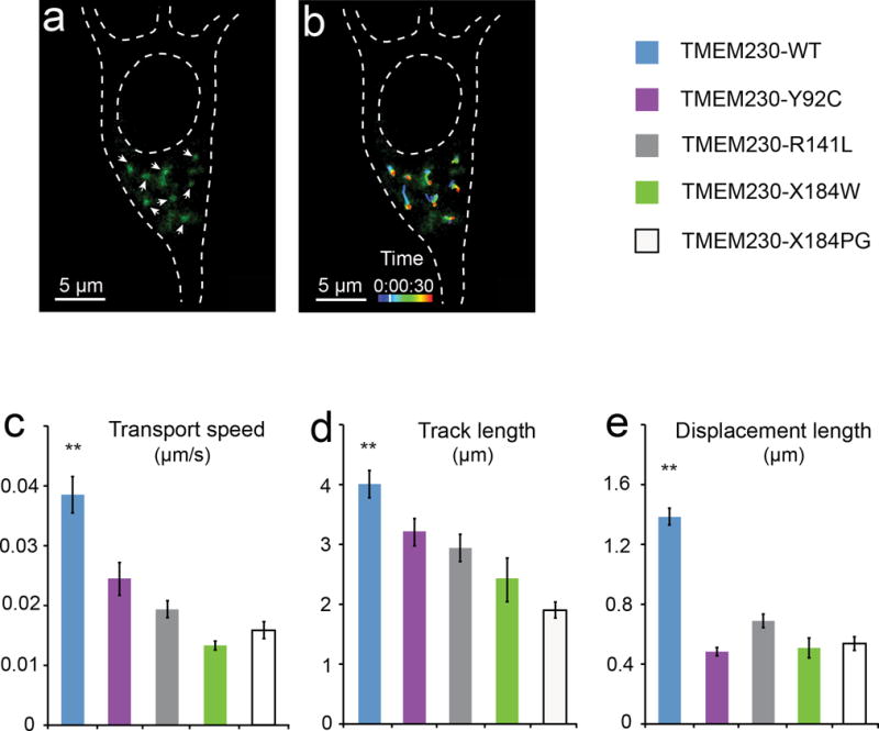
Impairment of synaptic vesicle trafficking by PD-linked mutant TMEM230. (a) Fluorescent image of synaptic vesicles in transfected primary neurons. Cultured mouse midbrain primary neurons were co-transfected with the GFP-tagged VAMP2 expressing vector (pEGFP-VAMP2) and each of the TMEM230 tag-free constructs in the pIRES2-ZsGreen1 vector. The pEGFP-VAMP2 was used to label the synaptic vesicles (Supplementary information). White arrowheads mark representative GFP-VAMP2-labeled vesicles quantified in panels b-e. (b) Movement track of the GFP-VAMP2-labeled vesicles. The trafficking of the vesicles in live primary neurons is monitored by confocal microscope and quantified using the Imaris software package. The movement tracks of eight representative vesicles from time-lapse live confocal images (30 frames with 5-second intervals) are shown, with pseudocolored movement tracks marking the position of vesicles at different time points during the 150 seconds. Images shown in panels (a) and (b) are taken at the time point marked on the pseudocolored time scale. (c-e) Quantification of the mean transport speed, track length and displacement length of GFP-VAMP2-labeled vesicles. Vesicles in neurons expressing PD-linked mutants (TMEM230-Y92C, TMEM230-R141L, TMEM230-X184W and TMEM230-X184PG) show impaired movement, as measured by transport speed (c), track length (d) and displacement length (e), when compared to the wild-type (TMEM230-WT). Data are from four independent experiments (1055 moving vesicles in 27 neurons). ** p < 0.01 (Oneway ANOVA test). Error bars, mean ± s.e.m.
