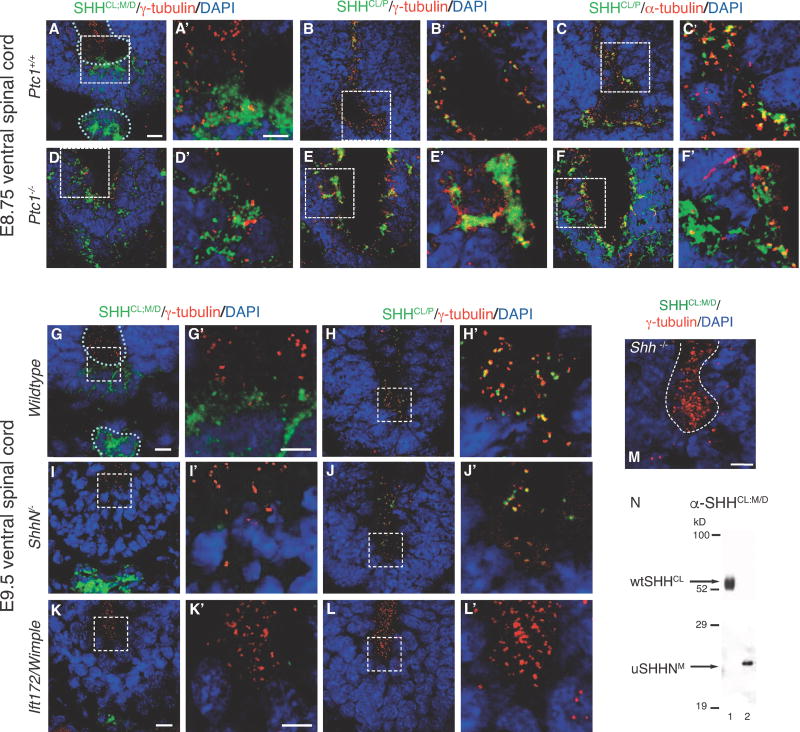
Figure 2. SHHCL/P localization in the embryonic ventral spinal cord of Ptc1−/−, ShhN/, and Ift172/Wimple mice.
SHHCl/P co-localization with cilia BBs is determined using immunofluorescence microscopy in embryonic ventral spinal cord sections of mice lacking PTC1, Shh cholesterol modification, and IFT172 mutation (Wimple). E8.75 ventral spinal cord: Shh+/+ (A–C, A′–C′), Ptcl−/− (D–F, D′–F′). E9.5 ventral spinal cord, Shh+/+ (G, H, G′, H′), ShhN/−, lacking the C-terminal cholesterol modification, (I,J, I′,J′), Ift172/Wimple (K, L, K′, L′). White dotted lines outline ventral spinal cord ventricles and notocord. Anti-SHH antibodies (α-SHHCL;P and α-SHHCL;M/D) are green. Anti-γ-tubulin detects cilia BBs in red (A, B, D, E, A′, B′, D′, E′, G–L, G′–L′, M)). Anti-a-tubulin detects cilia axonemes in red (C, C′, F, F′). Loss of PTC1 causes increased diffusion and aggregation of SHHCL/P (H, H′, I, I′), and diffusion of SHHCL;M/D (G, G′). α-SHHCL;M/D recognizes floorplate and notochord localized SHH, but not cilia BB associated SHH puncta. M. Anti-SHHCL;M/D (green) does not recognize targets in Shh−/− E9.5 ventral embryonic spinal cord, γ-tubulin (red). N. Western analysis using α-SHHCL;M/D recognizing an epitope shared by both wtSHHCL and uSHHN proteins. Scale Bars: 10μm (A–M), 5μm (A′–L′).

