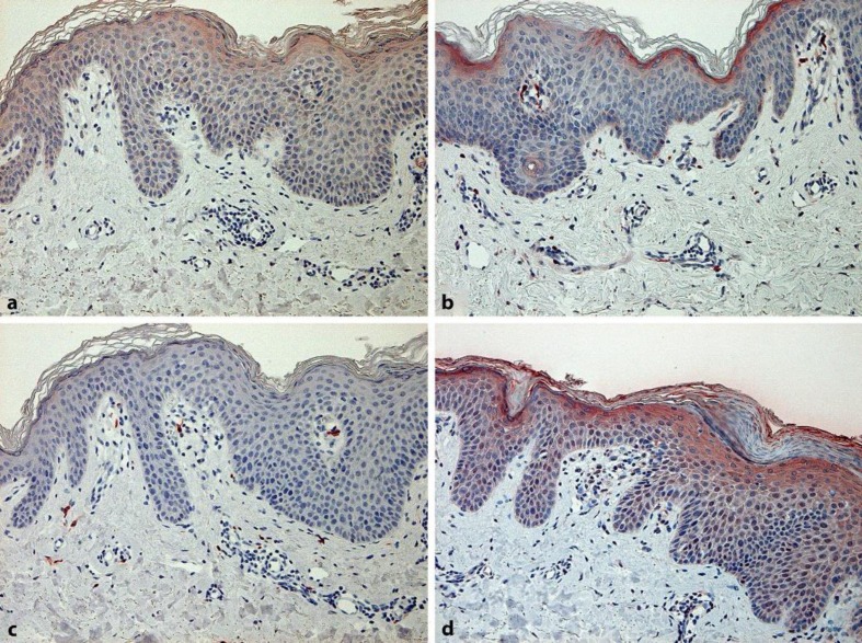
Fig. 3.
Immunohistochemical staining of the present case. Paraffin-embedded samples were deparaffinized and stained with anti-IL-36γ antibodies (a), anti-IL-36R antibodies (b), anti-IL-17 antibodies (c), and anti-IL-17R antibodies (d). The sections were developed with liquid permanent red. (Original magnification: ×200).

