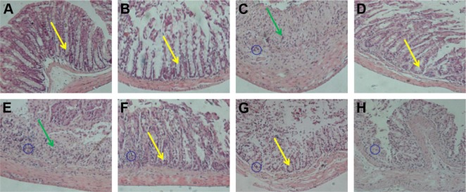
Figure 6.
Pathological analysis of colon tissues after distinct treatments using H&E staining assay, magnification =100×. (A) Normal control, (B) CA/BaSO4 microcapsule treatment, (C) DSS induction, (D) sulfasalazine treatment after DSS induction, (E) 0.6×10⁹ Bifidobacterium-loading CA/BaSO4 microcapsule treatment after DSS induction, (F) 1.2×10⁹ Bifidobacterium by microcapsule treatment after DSS induction, (G) 2.4×10⁹ Bifidobacterium by microcapsule treatment after DSS induction, (H) 2.4×10⁹ Bifidobacterium treatment after DSS induction. The blue circles denote infiltration of mononuclear, submucosal lymphoid hyperplasia, and extension around intestinal glands; yellow arrows denote the distortion of crypts and loss of goblet cells; the green arrows denote the severe mucosal damage in the colon.
Abbreviations: CA/BaSO4, chitosan-coated alginate microcapsule loaded with in situ synthesized barium sulfate; DSS, dextran sulfate sodium.

