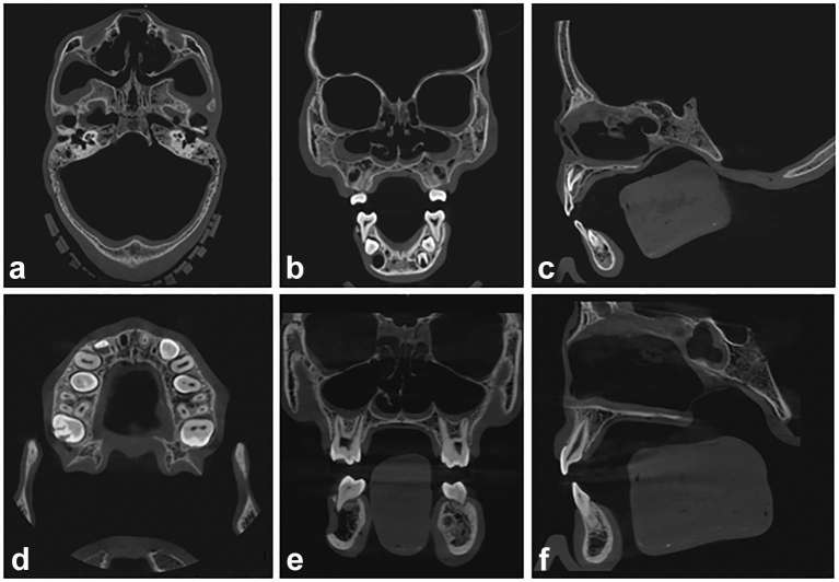
Figure 5.
CBCT images obtained on the CS9300 unit, from phantoms 1 (a–c; field of view 17 × 11 cm, 90 kV, 4 mA, voxel size 0.3 mm, 12 s) and 6 (d, e; a–c; field of view 10 × 10 cm, 90 kV, 4 mA, voxel size 0.18 mm, 8 s). The images show a perfect fit of the Mix-D over the bone surfaces and the smooth and continuous outline following the contour of the craniofacial structures. A slight overflow of Mix-D can be detected inside the cranium (a–c), maxillary sinus (b, e), sphenoid sinus (f), nasal cavity (b, c, e, f) and orbits (b, e). Axial (a, d) coronal (b, e) and sagittal views (c, f). CBCT, cone-beam CT.

