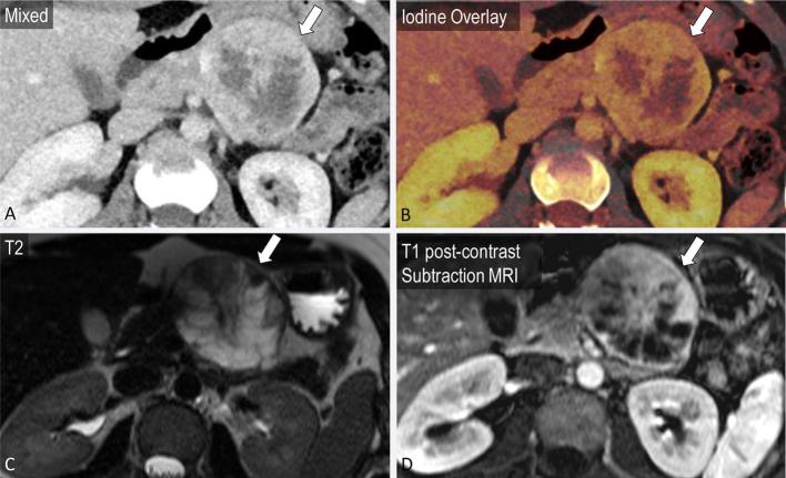
Figure 9.
A 25-year-old female with incidental finding after presentation for fall. A partially cystic mass in the tail of the pancreas is noted on mixed axial images from portal venous phase CT (a, block arrow). Iodine overlay images demonstrate nodular enhancement within the mass (b). T2 (c) and T1 post-contrast MRI images (d) confirm a cystic mass with areas of nodular enhancement corresponding to the iodine overlay images, most likely a pancreatic solid pseudopapillary epithelial neoplasm.

