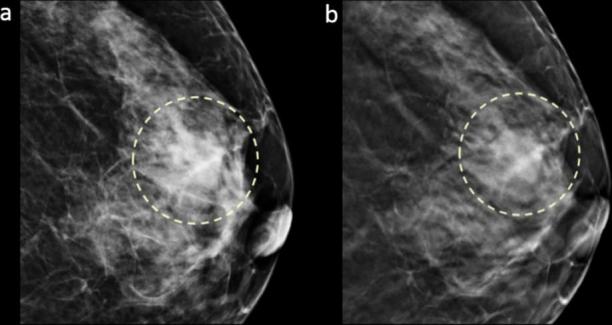
Figure 3.

Images in a 64-year-old female with a 2.0 cm, triple negative, histological Grade 3, invasive ductal carcinoma in dense breast (grade c). Craniocaudal DM (a) and DBT (b) images show a mass with oval shape and circumscribed margin in the left breast subareolar region that was detected by all three readers (detectability score 3).
