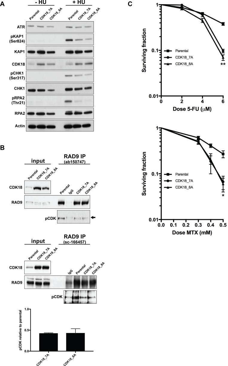
Figure 4. Heightened expression of endogenous CDK18 disrupts replication stress signalling and confers an increased sensitivity to replication stress-inducing agents.
(A) Representative western blots for the indicated proteins in untreated and HU-treated parental and CDK18 activation MDA-MB-231 clones. (B) Representative western blots of endogenous RAD9 IPs from parental and CDK18 activation clones using two separate human RAD9 mouse monoclonal antibodies (upper and middle panels as indicated). Equal amounts of immunoprecipitates were probed with either RAD9 (rabbit polyclonal) or phospho-CDK substrate (K/HpSP motif; pCDK) antibodies as indicated. Inputs demonstrate heightened CDK18 expression in the activation clones and comparable RAD9 expression. IgG was used as a negative control for non-specific binding which can be seen as a faint band migrating at a slightly higher molecular weight to RAD9 (black arrow). Levels of pCDK in the RAD9 IPs were quantified from these data by normalising to the relative amount of immunoprecipitated RAD9 within each cell population. The lower panel shows the quantified average pCDK levels in the indicated cell populations with their respective SEMs. (C) Clonogenic survival curves for 5-FU (left panel) and Methotrexate (MTX) treated (right panel) MDA-MB-231 cell lines as indicated. Data shown represents the mean derived from at least three independent experiments with their respective SEMs (*p ≤ 0.05 and **p ≤ 0.01 compared to parental MDA-MB-231 cells at the same drug dose).

