
Figure 6. uPAR knockout results in a decreased amount of Ki-67-positive Neuro 2A cells.
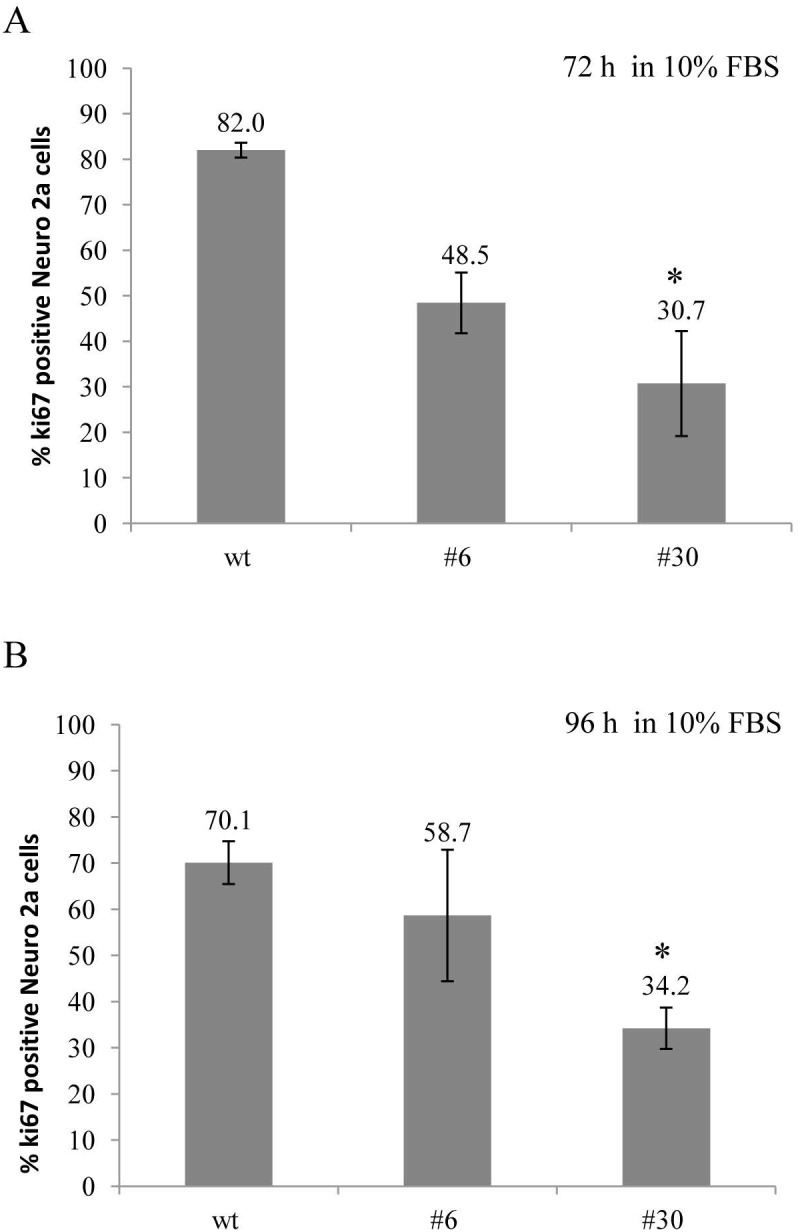
Ki-67 expression was assessed in uPAR-deficient clones (#6, #30) and in wt cells in standard culture media (10% FBS) after 72 and 96 hours. Cells were fixed, stained with antibody against Ki-67 and subjected to FACS analisys. (A) The bar chart depicts the percentage of Ki-67-positive Neuro 2A cells in each cell type after 72 hours; (B) The percentage of Ki-67-positive Neuro 2A cells in each cell type after 96 hours. The data are presented as Mean ± SD (n=4 wells per group), *p < 0.05 by ANOVA test.