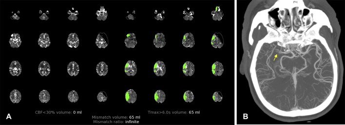
Figure 2.
(A) CT perfusion with RAPID software on arrival at hospital demonstrating 65 mL of maximum time to peak greater than 6 s (penumbra) and no core ischemic area. (B) CT angiography showing occlusion of the right middle cerebral artery (MCA; yellow arrow) and good collaterals (>50%) of the right MCA territory.

