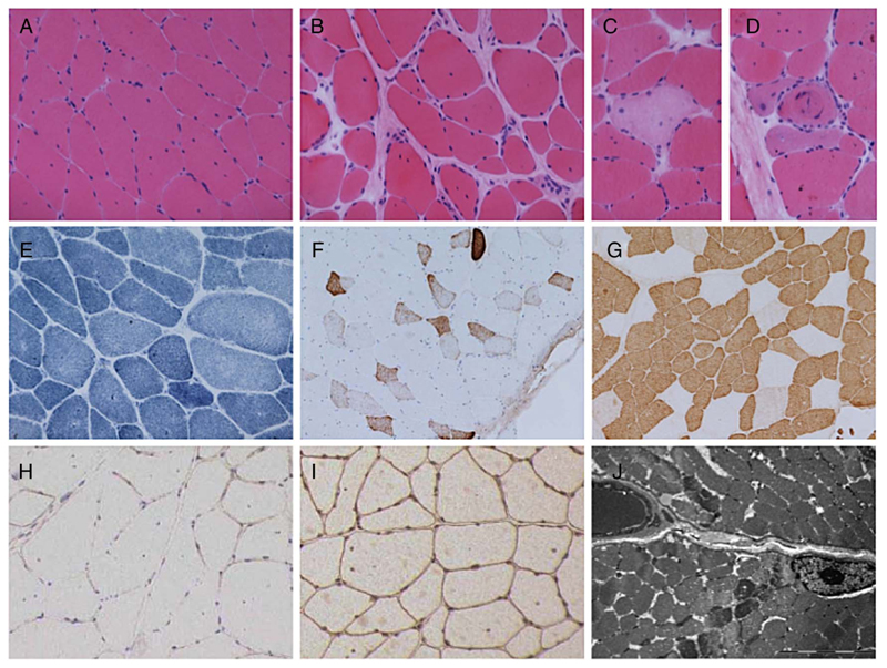
Figure 3.
Muscle biopsy. Muscle biopsy from quadriceps femoris in case 3 at age 25 years, showing variation in fibre size with occasional atrophic fibres and increase in internal nucleation with H&E staining (A). There is a focal increase in endomysial collagen (B), occasional necrotic fibres (C) and regenerating fibres (D). NADH staining shows mild disturbance of the internal architecture in a number of fibres (E). Numerous fibres express neonatal myosin (F). There is a pattern of slow fibre predominance with these being of generally small size (G). Immunohistochemical staining shows reduced labelling for α-dystroglycan (H) compared to β-dystroglycan (I). Ultrastructural examination shows lack of accumulation of granular material, rimmed vacuoles or tubular aggregates (J). Range of fibre size A-I=30–120 μm; bar in J=5 μm. NADH, nicotinamide adenine dinucleotide hydrogen.

