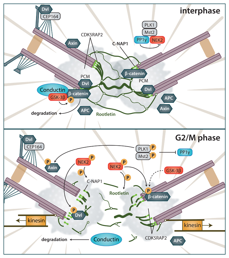
Figure 5. Effects of Wnt pathway components on the centrosome separation.
(Upper) Interhase cells. Centrosomes are connected by a linker that consists of fibrous Rootletin and is anchored to the proximal sides of centrioles by C-NAP1. During interphase, centrosome cohesion is maintained by action of Axin2/Conductin and GSK-3β by phosphorylation of β-catenin. DVL in ciliated cells promote basal body docking, via interaction with CEP164 and other proteins. Axin and APC can be also found at centrosome, probably due to their microtubule interactions. NEK2 is kept inactive in complex with PLK1/MST2/PP1γ. (Lower) G2/M phase. Phosphorylation cascade activates NEK2, which subsequently acts on several targets. β-catenin is protected from degradation by NEK2 phosphorylation, and promotes centrosome separation; Axin2/Conductin is degraded by proteasome. Concurrently, phosphorylation of DVL and C-NAP1 by NEK2 leads to the increase in their overall negative charge and subsequent release from centrioles. Centrosomes can be subsequently pulled apart. Axin is phosphorylated by PLK1, which affects microtubule dynamics during spindle formation. For details and references see text.

