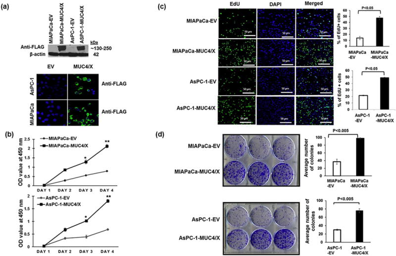
Fig. 2.

Functional implications of overexpression of the MUC4/X on PC cell proliferation and colony formation. (a) MUC4/X was cloned into the N-terminus FLAG-tagged, and the C-terminus HA-tagged p3XFLAG-CMV™-9 vector followed by stable transfection into WT-MUC4 non-expression PC cell lines MIAPaCa and AsPC-1. (Upper panel) Immunoblot analyses using anti-FLAG antibody was performed to assess the expression of MUC4/X. MUC4/X overexpression was observed in stably transfected MIAPaCa-MUC4/X and AsPC-1-MUC4/X cells whereas no expression was detected in empty vector (EV) transfected cells. (Lower Panel) Immunofluorescence analyses for MUC4/X using anti-FLAG antibody showed expression of MUC4/X in MIAPaCa-MUC4/X, and AsPC-1-MUC4/X PC cells while no expression was observed in vector alone transfected cells. (b) MTT assay was performed to assess the impact of MUC4/X overexpression on cellular proliferation. Significant higher cell proliferation in MIAPaCa-MUC4/X and AsPC-1-MUC4/X cell lines were observed as compared to vector transfected control cells at 3rd and 4th days (MIAPaCa-EV/AsPC-1-EV) (**p < 0.01, *p < 0.05). Line diagram represents the OD value at 450 nm (mean ± SD, n = 3). (c) EdU cell proliferation assay was performed to assess the impact of MUC4/X overexpression on PC cell proliferation. MUC4/X-OE cells had a higher number of EdU positive proliferating (green fluorescent) cells as compared to control vector transfected cells (p < 0.05) after 24 h of incubation with EdU. Nuclei staining with DAPI represents total number of cells. Accompanying bar diagram demonstrates the quantitative measurement for percentages of EdU positive green fluorescent cells (mean ± SD, n = 3). (d) Colony forming assay suggested MUC4/X-transfected MIAPaCa and AsPC-1 cells formed significantly greater number of colonies than the control cells (p < 0.005). The corresponding bar diagram represents quantitative analysis of an average number of colonies (mean ± SD, n = 3) observed per well of six-well plates. The two-tailed, unpaired t-test analyses was used to determine significance between two groups. These results suggest MUC4/X overexpression promotes cell proliferation and colony formation.
