Figure 2.
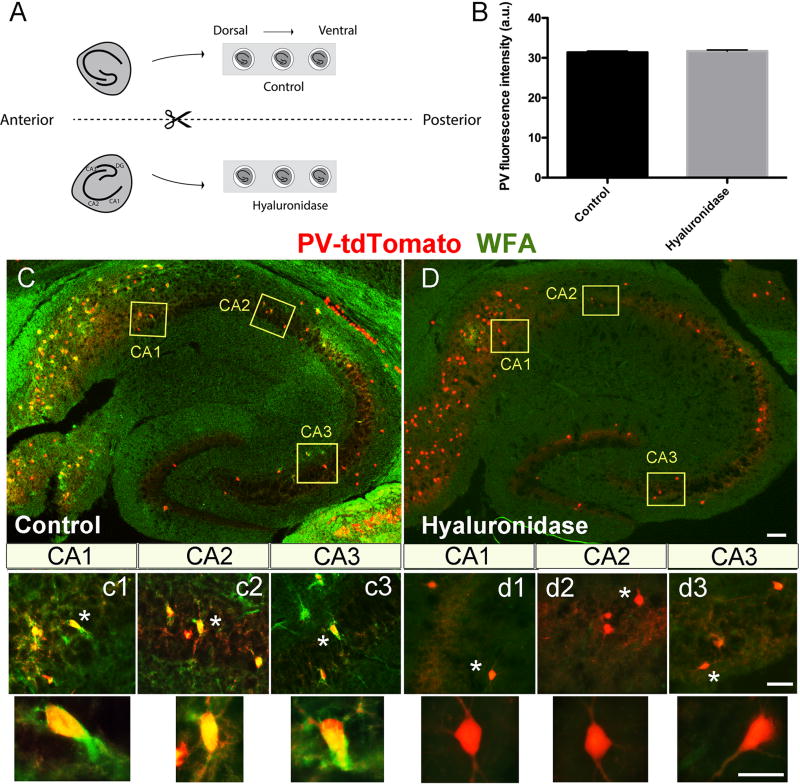
Horizontal slice preparation and staining. Horizontal slices were prepared and bisected, with matched hemisections placed in one of two recovery solutions (ACSF or ACSF with hyaluronidase). Following a 2h recovery period, hemislices were used for additional studies. A schematic of the set up is shown in 2A, and representative PV fluorescence in cells from control (n=1392; 6 fields) and hyaluronidase (n=927; 5 fields) treated slices, prepared from PVtd-Tomato mice, is shown in 2B. No significant difference was detected. In 2C and D, we show representative immunostaining of a hippocampal slice from a PVtd-Tomato mouse with WFA. Layers of CA1, CA2, and CA3 areas indicated by yellow rectangles are shown magnified in c1–c3 for control slices, and d1–d3 for hyaluronidase treated slices, respectively. WFA positive PV expressing cells can be appreciated in control but not hyaluronidase treatment conditions. Scale bars: 100 µm (C, D), 50 µm (c1–c3, d1–d3), and 20 µm for high magnification single neurons.

