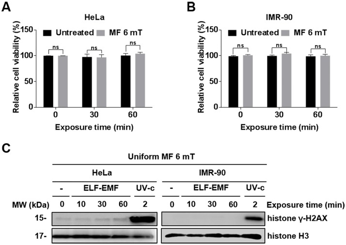
Fig 2. Cellular effects of a single exposure to a uniform EMF of 6 mT.
(A, B) HeLa and IMR-90 cells were exposed to an EMF of 6 mT for 0, 30, and 60 min. Cell viability was measured by MTT assays and evaluated as a percentage relative to the viability of unexposed cells (0 min). Values are presented as the mean ± SD (n = 3) and P-values were determined by two-way ANOVA with the Bonferroni correction. Values of *P < 0.05, **P < 0.01, ***P < 0.001, and ****P < 0.0001 were considered statistically significant, and P > 0.05 was considered statistically not significant (ns). (C) γ-H2AX was assessed by western blot analysis. Histone H3 was used as a loading control and cells exposed to ultraviolet (UV) (100 J/m2) were used as positive controls for DNA damage.

