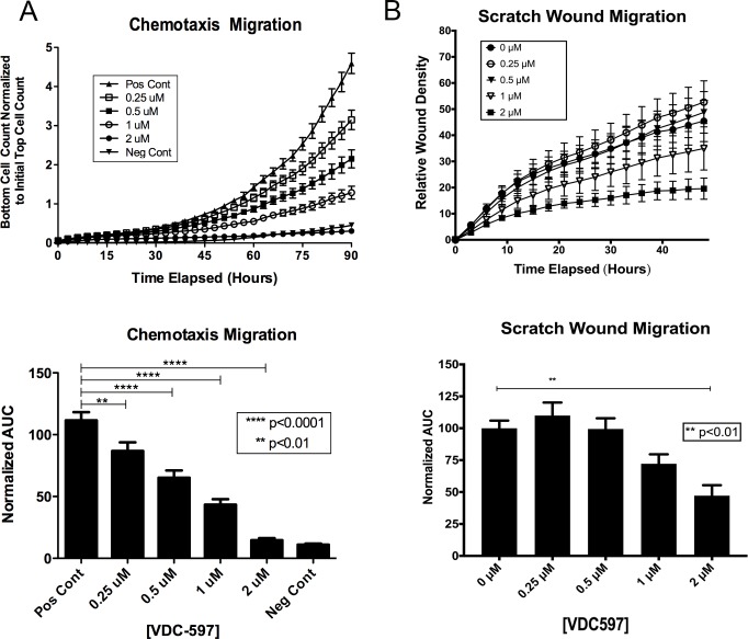
Fig 6. VDC-597 inhibits canine hemangiosarcoma cell migration.
A. DEN-HSA was plated in the top chamber of a specialized 96-well chemotaxis migration plate in either C/0.1 EMEM or C/0.1 EMEM with various concentrations of VDC-597. C/20 EMEM or C/20 EMEM with identical various concentrations of VDC-597 of the top chamber was placed in the bottom chamber. Migration of the cells through the micropore membrane was assessed using the Incucyte Live Cell Imaging System over 90 hours. Area under the curve for each condition is expressed. Asterisks indicate P values less than 0.05. B. DEN-HSA was plated and allowed to grow to confluency in a specialized 96-well scratch wound assay plate. A uniform defect (“scratch”) was made through the confluent layer of cells and mitomycin C-containing C/10 EMEM with various concentrations of VDC-597 was added. Relative wound density was assessed using the Incucyte Live Imaging System over 48 hours. Area under the curve for each condition is expressed. Asterisks indicate P values less than 0.05. Error bars indicate standard error measurements.

