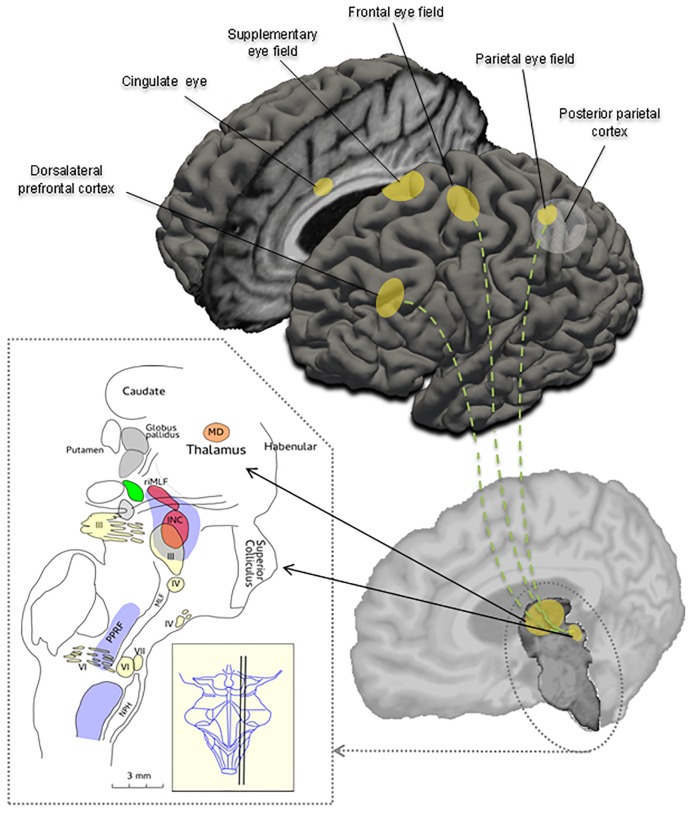
Fig 1. Schematic overview of important cortical regions and brainstem areas involved in the control of eye movements.
The sagittal cross section of the brainstem is showing the three oculomotor nuclei (III, IV, VI), the nucleus prepositus hypoglossi (NPH), the interstitial nucleus of Cajal (INC), the superior colliculus, the paramedian pontine reticular formation (PPRF), the medial longitudinal fasciculus (MLF) and the mediadorsal nucleus (MD) in the thalamus. The nucleus raphe interpositus (not shown) lies close to the midline, at the level of the abducens nucleus (VI) [modified from Petzold A, Paine M, Faldon M, Riordan-Eva P, Bronstein AM, Gresty MA, Plant GT. Synchronised paroxysmal ocular tilt reaction and limb dystonia. Neuroophthalmology 2009;33:217–236.].

