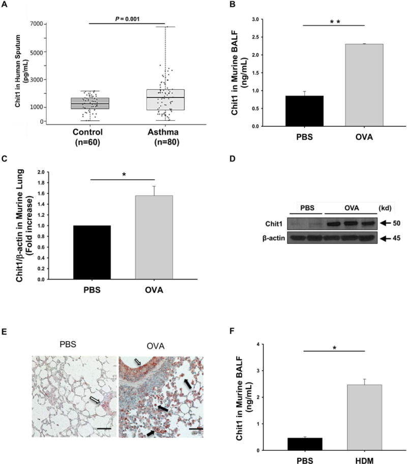
Figure 1. Chit1 expression in childhood asthma and allergen-induced airway inflammation mice.

(A) Chit1 levels in supernatants of induced sputum from patients with normal control subjects (n = 60) and asthma (n = 80). (B-E) WT and Chit1−/− mice were sensitized with OVA/Alum and challenged with OVA. 24h After the last OVA challenge, the levels of Chit1 protein and mRNA expression were assessed via (B) ELISA and (C) real-time PCR and (D) Western blot. (E) Representative immunohistochemistry on lung sections with vehicle (PBS) or OVA challenge (solid arrows, alveolar macrophages; open arrow, airway epithelial cells). (F) WT and Chit1−/− mice were sensitized with house dust mite (HDM)/Alum and challenged with HDM. The level of Chit1 in bronchoalveolar lavage (BAL) fluids was assessed using ELISA. The values in panels B-D, F represent the triplicate evaluations in a minimum of 5 mice each group. Panels E is representative photograph of minimum 5 mice each group. Scale bars in E; 100 μm, respectively. *p<0.05, **p<0.01.by Pearson test and student’s t-test.
