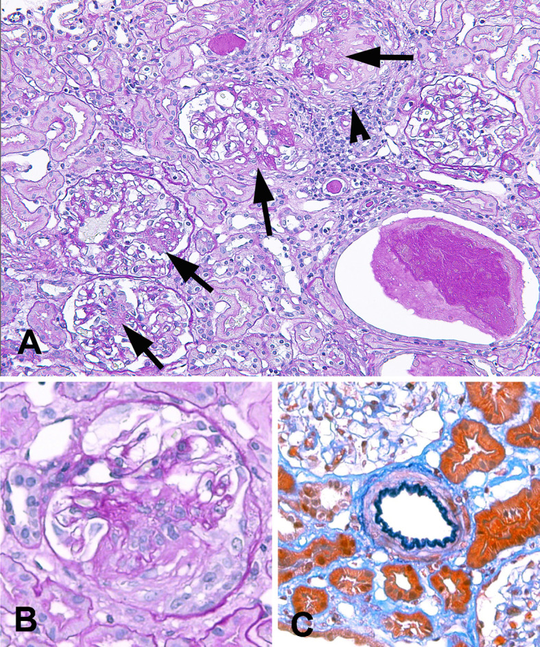
Figure 1.

Light microscopic findings of the kidney biopsy. A. Light micrograph shows glomeruli with focal segmental necrotizing lesions (arrows). There are proteinaceous cast formations and mononuclear cell infiltration around a glomerulus with extensive dissolution of the Bowman’s capsule (arrow head). Tubulointerstitial nephritis with lymphoplasmacytic infiltration is not observed. (PAS staining) ×200. B. A glomerulus with fibrinoid necrosis and fibrocellular crescent formation (PAS staining) ×400. C. A small artery shows no vasculitis (Elastica-Masson staining) ×400. PAS: periodic acid-Schiff
