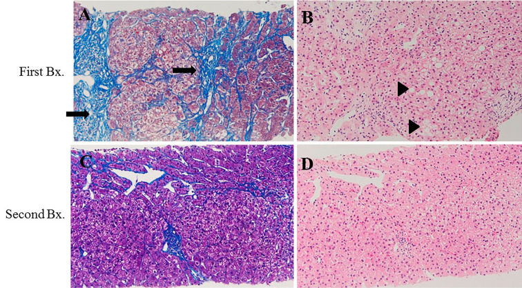
Figure 3.
Liver biopsy. First biopsy (A) (B) showed steatohepatitis, which was classified as stage 3-4; grade 1 according to Brunt’s classification. The arrow indicates expanded fibrosis. The triangle indicates steatosis. After 6 months, a second biopsy was performed (C) (D). The fibrosis stage was found to have decreased to stage 2-3, and the steatosis and ballooning hepatocytes were diminished. (A) and (C), Mallory Azan staining. (B) and (D), Hematoxylin and Eosin staining.

