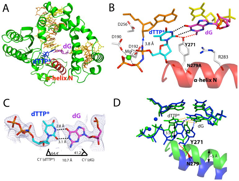
Figure 2.
Ternary structure of Asn279Ala polβ in complex with the dG:dTTP* mismatch in the presence of active-site Mg2+ (PDB ID 4Z6D). (A) Overall structure of Asn279Ala polβ with the templating dG paired with nonhydrolyzable dTMPNPP (dTTP*). The incoming dTTP* is shown in blue. The templating dG is shown in magenta, whereas the rest of templating strand bases are shown in yellow. The upstream and downstream primers are shown in orange. The α-helix N is shown in red. (B) Close-up view of the active site of the Asn279Ala dG:dTTP-Mg2+ ternary structure. The catalytic aspartates and amino acid residues involved in minor groove edge recognition are indicated. (C) Base pairing between the templating dG and the incoming dTTP*. The C1′ distance and λ angles are shown. A 2Fo-Fc map contoured at 1σ around dG:dTTP* is shown. Distances for O6(dTTP*)-O6(dG), N1(dTTP*)-N1(dG), and O2(dTTP*)-N2(dG) are 3.8Å, 3.9Å, and 3.9Å, respectively, indicating a wobble dG:dTTP* base pair. (D) Comparison of the active site of the Asn279Ala polβ-dG:dTTP*-Mg2+ structure (shown in green) with that of the published wild-type polβ-dG:dTTP*-Mg2+ structure (shown in blue, PDB ID 4PGQ) (19).

