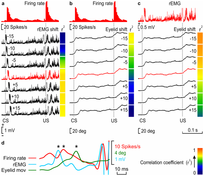
Figure 5.
Timing correlation between firing rate, rEMG, and lower-eyelid position for CRs collected during classical eyeblink conditioning. We calculated the coefficient of determination (r2) between the firing rate of type A neurons and the rEMG (a), the firing rate and eyelid position (b), and the rEMG and eyelid position (c) in 10 averaged trials. (a) A representation of r2 values between the firing rate of IPN neurons and the corresponding rEMG (red trace), when the rEMG was shifted −15 ms, −10 ms, −5 ms, +5 ms, +10 ms, and +15 ms (black traces) from the original timing (0 ms, red trace) recorded during a conditioning session of a WT trained mouse during a delayed conditioning paradigm. We compared regions of the CS-(US −20 ms) interval and delimited each one by turning points automatically detected in the firing rate. For each comparison, the collected r2 value is represented by a colored square to the right of each rEMG recording. (b) Same correlation was calculated for neuron firing rate vs. eyelid position. (c) Same correlation is calculated for rEMG vs. eyelid position. The calibration for color gradient values is shown at the bottom right in (d). Higher r2 values corresponding to negative-shifted recordings indicate a delay with respect to the correlated recording (in a and b, the firing rate, and in c, the rEMG), whereas higher r2 values corresponding to positive-shifted recordings indicate an advance with respect to the correlated recording. (d) Superimposition of smoothed recordings of the firing rate, rEMG, and eyelid movement, corresponding to the last 80 ms of the CS-US interval of the unshifted recordings shown from (a) to (c), showing the timing appearance of bursts of the CR (*). Note that, whereas eyelid movement is delayed with respect to the firing rate (b) and, obviously, with respect to the rEMG (c), interestingly the rEMG is advanced with respect to the firing rate (a).

