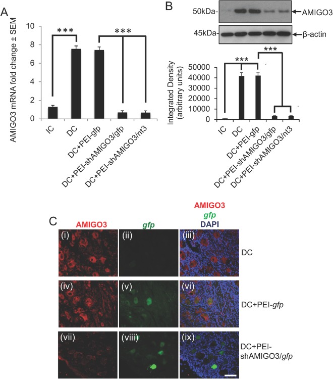
Figure 3.
AMIGO3 levels are suppressed in DRGN after injection of PEI transduced plasmids encoding shAMIGO3. (A) Low levels of AMIGO3 mRNA in IC increased significantly after DC injury (P < 0.0001) and remained high in the DC + PEI-gfp group. AMIGO3 mRNA levels reduced significantly in both DC + PEI/shAMIGO3/gfp and DC + PEI-shAMIGO3/nt3 groups (P < 0.0001). (B) Western blot and subsequent densitometry for AMIGO3 protein reflected the changes in mRNA. β-actin was used as a loading control (full scans shown). (C) Immunohistochemistry for AMIGO3 showed high levels of AMIGO3 (red) in DRGN after DC (C(i)), which were gfp− (C(ii)). AMIGO3 levels remained high in DC + PEI-gfp groups (C(iv)), with gfp expression (green) in some DRGN (C(v)). AMIGO3 levels were suppressed in DRGN from DC + PEI-shAMIGO3/gfp groups (C(vii)), with gfp expression in some DRGN (C(viii)). (C(iii)), (C(vi)) and (C(ix)) are merged images showing AMIGO3 (red), gfp (green) and DAPI stained nuclei (blue). Scale bar = 100 μm. ***P < 0.0001, ANOVA.

