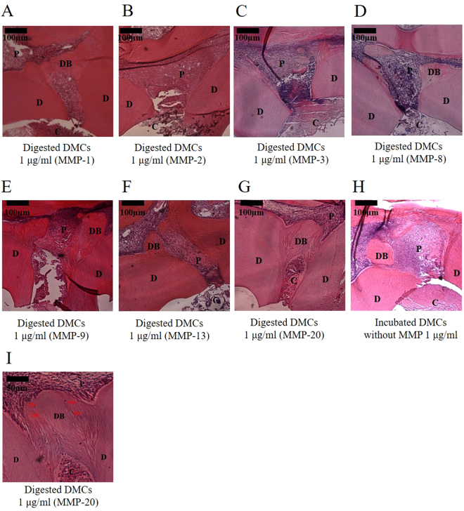
Figure 8.
Histological images (100×) of tertiary dentin formation 28 days after direct pulp capping using 1 µg/ml of dentin matrix components (DMCs) treated with matrix metalloproteinase (MMP)-1 (A), MMP-2 (B), MMP-3 (C), MMP-8 (D), MMP-9 (E), MMP-13 (F), MMP-20 (G), or incubated DMCs without MMP (H). Higher magnification image (200×) of 1 µg/ml of DMCs digested with MMP-20 (I). 1 µg/ml of DMCs treated with MMP-1, -9, -13, or -20 facilitated hard tissue repair, compared with 1 µg/ml of DMCs treated with other MMPs or incubated DMCs without MMP, based on the rate of coverage of the exposed pulp by tertiary dentin (p < 0.05) (see online Supplementary Tables 1 and 2). In particular, 1 µg/ml of DMCs treated with MMP-20 induced a significantly higher condense and more regular tubular structure. Significant differences were not observed with lower concentrations (0.01–0.1 µg/ml) of the above-described MMPs. These data represent six independent experiments. Arrows indicate dentinal tubules. C = cavity, D = dentin, DB = Tertiary dentin bridge, P = pulp.

