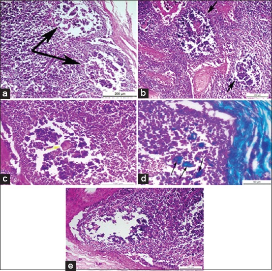
Figure-2.

(a) Photomicrograph of a buffalo lymph node showing necrotic depleted and rarified sub-capsular lymphoid follicles, two black arrows (H and E, Bar=200 µm), (b) photomicrograph of buffalo lymph node showing multiple, necrotic, depleted, and rarified deep cortical lymphoid follicles near to corticomedullary junction, two black arrows (H and E, Bar=200 µm), (c) photomicrograph of a buffalo lymph node showing single eosinophilic structure less mass replacing the necrotic area of lymphoid follicle, yellow arrow (H and E, Bar=100 µm), (d) photomicrograph of a buffalo lymph node showing multiple green colored structure less masses of collagen, three black arrows (Masson’s trichrome, Bar=50 µm), (e) photomicrograph of a buffalo lymph node showing total lack of the germinal center of the lymphoid follicle (H and E, Bar=100 µm).
