Figure-3.

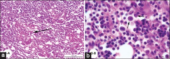
(a) Photomicrograph of a buffalo lymph node showing focal macrophage cell granulomatous reaction surrounds a focal area of necrosis (H and E; Bar=100 µm), (b) Photomicrograph of a buffalo lymph node showing diffuse macrophage cell granulomatous reaction, black arrows and presence of few numbers of neutrophils, yellow arrow (H and E; Bar=20 µm).
