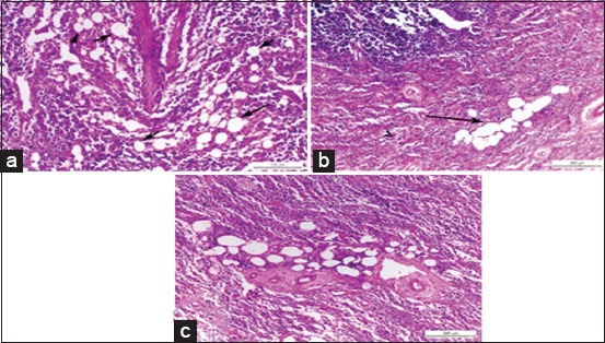
Figure-4.

(a) Photomicrograph of a buffalo lymph node showing multiple discrete variables sized fat globules in corticomedullary junction, black arrows (H and E, Bar=100 µm), (b) photomicrograph of a buffalo lymph node showing coalescence of fat globules into a single large globule, black arrow (H and E, Bar=200 µm), (c) photomicrograph of a buffalo lymph node showing multiple fat globules around blood vessels in the deep medulla (H and E, Bar=200 µm).
