Fig. 3.
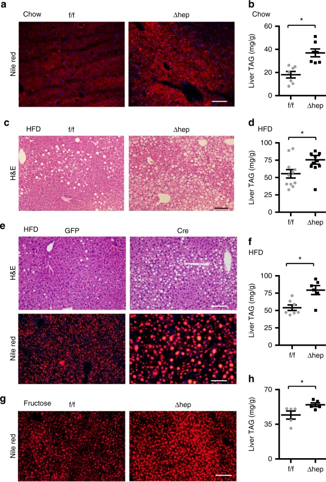
Hepatocyte-specific deletion of Snail1 promotes NAFLD. a, b Snail1flox/flox (n = 7) and Snail1Δhep (n = 7) males (18 weeks) were fed a normal chow diet. Livers were isolated under non-fasted conditions. a Representative Nile red staining of liver sections. b Liver TAG levels (normalized to liver weight). c, d Snail1flox/flox (n = 11) and Snail1Δhep (n = 11) male littermates were fed a HFD for 10 weeks. c Representative H&E staining of liver sections. d Liver TAG levels (normalized to liver weight). e, f Snail1flox/flox males were fed a HFD for 6 weeks and transduced with AAV-TBG-GFP (n = 7) or AAV-TBG-Cre (n = 6) vectors for 4 weeks. e Representative H&E or Nile red staining of liver sections. f Liver TAG levels (normalized to liver weight). g, h Snail1flox/flox (n = 5) and Snail1Δhep (n = 5) males were fed a fructose diet for 10 weeks. Scale bars: 100 µm. Data are represented as mean ± SEM. *p < 0.05, two-tailed unpaired Student’s t test

