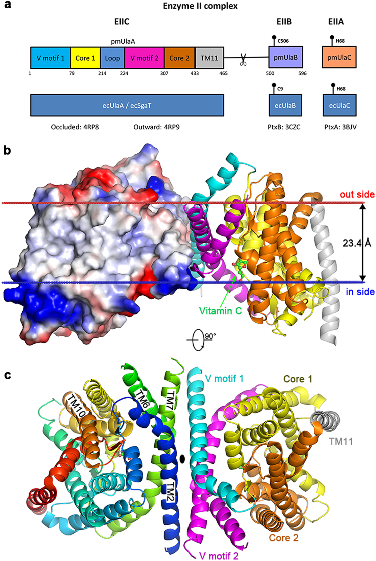
Fig. 1. Domain composition of l-ascorbate PTS enzyme II complex and overall structure of pmUlaA.
a In the wild type PTS EII complex from Pasteurella multocida, pmUlaB is fused C-terminal to pmUlaA to form a single peptide. His68 of UlaC accepts the phosphate group transferred from PEP, EI, HPr sequentially, and then gives it to Cys506/Cys9 of UlaB. Ultimately, UlaB transfers its phosphate group to the l-ascorbate. Residue 475, indicated by the scissor, was the proteolytic site with trypsin. b The structure of the pmUlaA homodimer is shown in cartoon and electron potential surface representations as viewed within the plane of the membrane. pmUlaA is spatially organized into V motif 1 (cyan), core 1 (yellow), V motif 2 (magenta), core 2 (orange) and TM11 (gray) subdomains. Vitamin C is shown in ball-and-stick representation. The spatial position of pmUlaA inside the lipid bilayer is predicted by the PPM server24. c View from the extracellular side of the pmUlaA homodimer. One protomer is shown in rainbow colors

