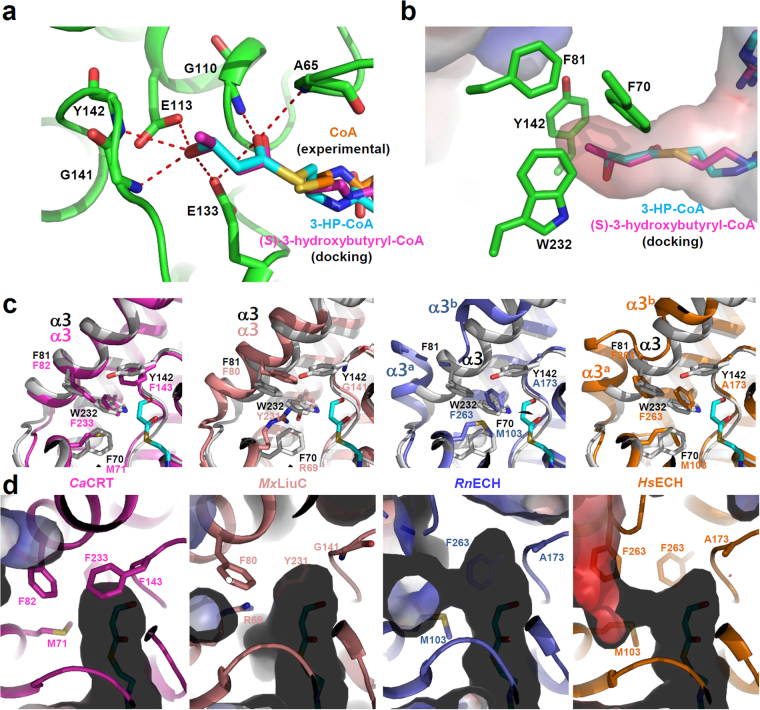
Figure 4.
Substrate specificity of Ms3HPCD. (a) 3-HP- and (S)-3-hydroxybutyryl-moiety binding mode of Ms3HPCD. The Ms3HPCD structure is shown as a cartoon diagram with a green color. The residues involved in the formation of the 3-HP binding pocket are shown as stick models and labeled appropriately. The bound CoA, simulated 3-HP-CoA, and simulated (S)-3-hydroxybutyryl-CoA are presented with stick models with colors of cyan, orange, and magenta, respectively. Hydrogen bonds involved in the 3-HP and (S)-3-hydroxybutyryl-moiety binding are shown as red-colored dotted lines. (b) Electrostatic potential surface model of the 3-HP binding pocket of Ms3HPCD. The Ms3HPCD structure is shown as an electrostatic potential surface presentation. The simulated 3-HP-CoA and (S)-3-hydroxybutyryl-CoA are presented by a stick model with cyan and magenta colors. The residues involved in the formation of the 3-HP binding pocket are shown as stick models with a green color. (c) Structural comparison of Ms3HPCD with CaCRT, MxLiuC, RnECH, and HsECH. The structure of Ms3HPCD is superposed with each of those of CaCRT, MxLiuC, RnECH, and HsECH. The structure of Ms3HPCD is shown with a gray color, and those of CaCRT, MxLiuC, RnECH, and HsECH are with colors of magenta, salmon, light-blue, and orange, respectively. The residues involved in constitution of the enoyl-binding pocket are shown as stick models. (d) Electrostatic potential surface model of the 3-HP binding pocket of other ECHs. The structures of CaCRT, MxLiuC, RnECH, and HsECH are shown as cartoon models and electrostatic potential surface presentations with color scheme same as in (c). The residues involved in the formation of the enoyl-binding pocket are shown as stick models.

