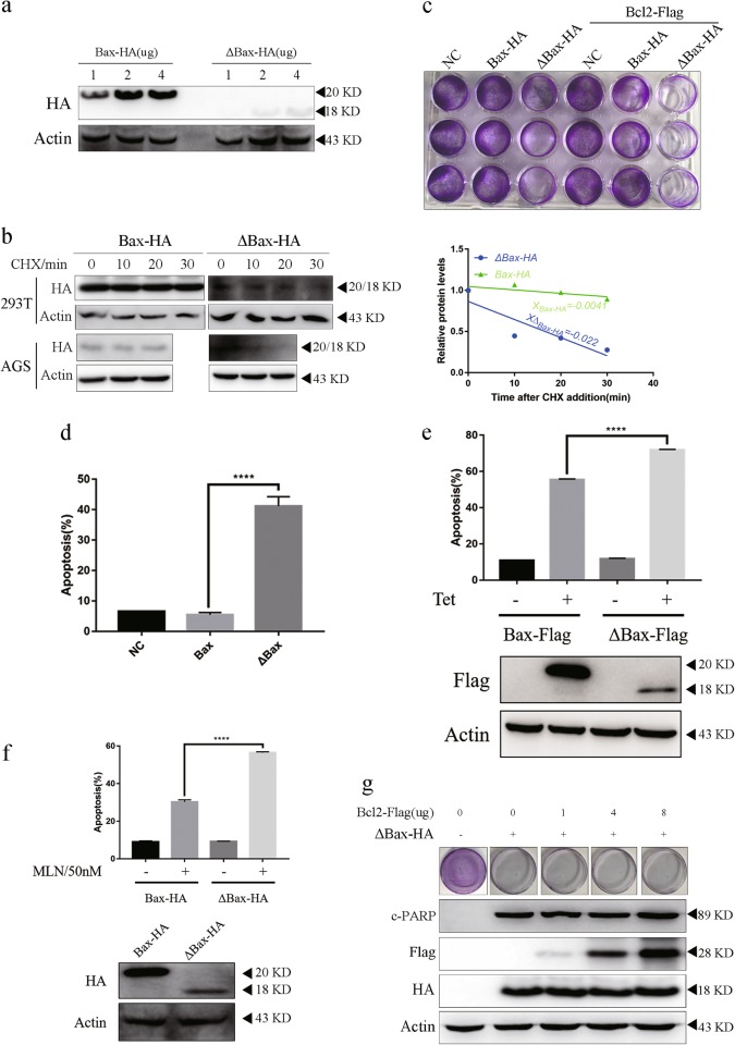
Fig. 5. Bcl-2 cannot block apoptosis induced by c-Bax.
a Differential expression of Bax-HA and ΔBax-HA. AGS cells were transfected with 1 µg, 2 µg, or 4 µg of either Bax-HA or ΔBax-HA for 24 h and subjected to immunoblotting for protein expression. b Short-lived ΔBax-HA than Bax-HA. CHX (50 ug/ml) was added to 293 T or AGS cells transfected with plasmid encoding Bax-HA or ΔBax-HA prior to treatment and cultured for 24 h. Equivalent lysates from the cells harvested at the indicated time points were immunoblotted for indicated protein expression. c, d, g Bcl-2 did not decrease apoptosis induced by ΔBax-HA. 1 µg Bax-HA or 4 µg ΔBax-HA plasmid was transfected with 4 µg Bcl-2-Flag (c) or 4 µg ΔBax-HA and indicated quality of Bcl-2-Flag plasmid, then cultured for 24 h and AGS subjected to crystal violet staining or immunoblot (c, g). AGS cells were treated with 1 µg Bax-HA or 4 µg ΔBax-HA plasmid (d) and subjected to Annexin V/propidium iodide double staining and FCM to detect apoptosis. e, f Cleaved Bax was a stronger apoptosis inducer. e The tet-on system was constructed and protein expression was induced by 10uM Tetracyclines for 24 h in AGS. Apoptosis level and protein level was detected by FCM and immunoblot. f The AGS stable cell line was constructed and incubated with 50 nM MLN8237 for 72 h. Apoptosis level and protein level was detected by FCM and immunoblot. Error bars represent SD from three independent experiments. This immunoblot is representative of three independent experiments. Asterisk (*) indicates a significant difference. ****P < 0.0001 two-tailed Student’s t test

