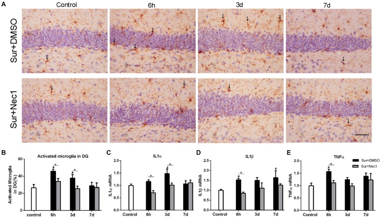
Figure 2.
The inhibition of RIPK1 by Nec-1 alleviates surgery-induced neuroinflammation in D-Gal-induced aged mice. (A) Representative images of Iba-1 staining (yellow) in the dentate gyrus. The typical activated microglial cells labeled by Iba-1 staining were indicated with arrows. Bar = 50 μm. (B) The percentage of Iba-1 immunopositive activated microglia was counted in dentate gyrus. Panels (C–E) represent qPCR analysis of mRNA levels of inflammatory factors IL-1α, IL-1β and TNF-α in the hippocampus. Data were expressed as the mean ± SEM. #P < 0.05, compared with control group; *P < 0.05, in comparison between Sur+DMSO and Sur+Nec1 groups by two-way ANOVA followed by Tukey’s multiple comparisons test (n = 6 per time point per group).

