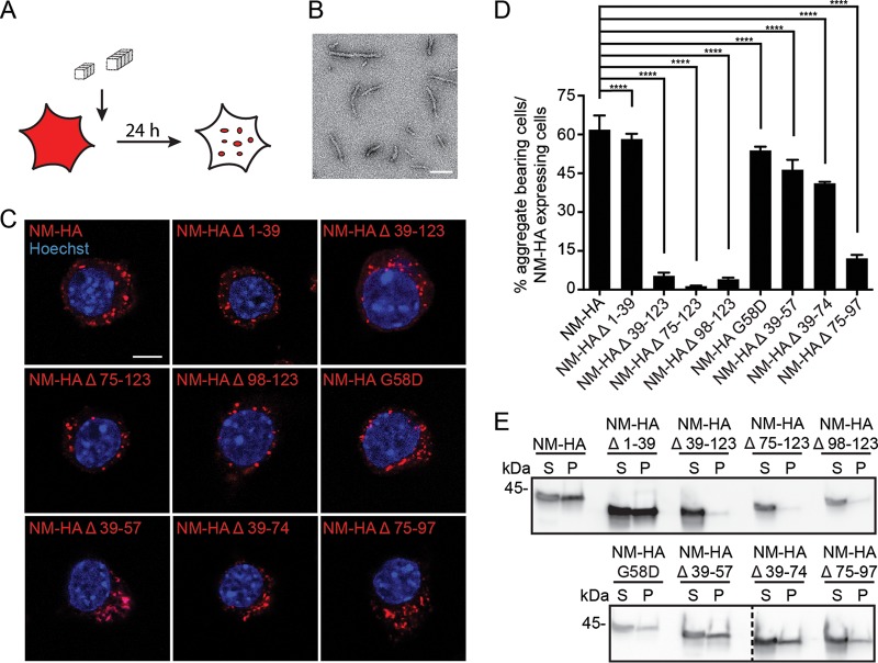
FIG 4.
Template-assisted aggregate induction by fibrillized untagged recombinant NM or endogenous NM-GFP prions. (A) Experimental setup to study the propensity of the NM mutants to form aggregates upon exposure to in vitro-fibrillized recombinant NM. (B) Electron microscopy image of untagged recombinant NM fibrils. Scale bar, 100 nm. (C) N2a cell populations exposed to 10 μM untagged recombinant NM fibrils (monomer concentration) for 24 h. Ectopically expressed wild-type and mutant NM were detected using MAb anti-HA (red), and nuclei were stained with Hoechst (blue). Scale bar, 5 μm. (D) Percentage of cells harboring NM-HA aggregates upon induction with 10 μM untagged recombinant NM fibrils (monomer concentration). Bars represent mean values ± SEM (three independent induction experiments; n = 3). At least 12,500 cells per cell population were imaged. Statistical analysis was done using the Cochran-Mantel-Haenszel test (****, P ≤ 0.0001). (E) Sedimentation assay of cell lysates from N2a bulk populations exposed to 10 μM fibrillized untagged recombinant NM (monomer concentration). Cells were harvested 24 h after aggregate induction. Ectopically expressed NM was detected using MAb anti-HA. S, supernatant; P, pellet. Additional lanes were excised for presentation purposes.

