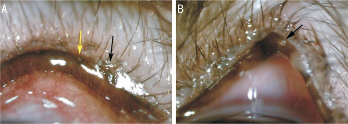
Figure 4. Morphological changes in the eyelid margin by slit-lamp observation.
A: Meibomian gland orifices were blocked after generating the model (black arrows). The wound was confined to the orifices. Yellow arrows show the meibomian gland orifices before generating the model; B: Black arrows show meibomian gland openings with hoary secretions blocked at the fourth week.

