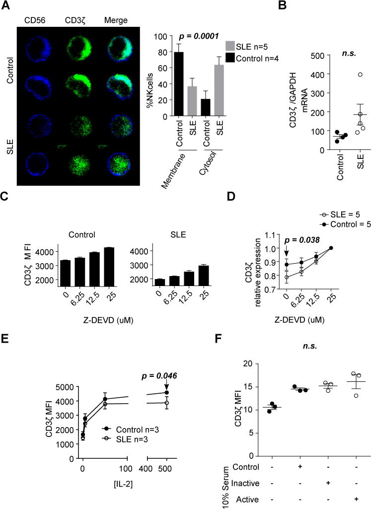
Figure 2. Caspase 3 inhibition preserves CD3ζ levels in NK cells from patients with SLE.

(A) NK cells from healthy donors and patients with SLE were stained with anti-CD56 (Blue) and anti-CD3ζ (Green) and visualized by confocal microscopy. Representative images are shown. The graph shows the percentage of cells with CD3ζ staining in the membrane or cytosol (mean ± SEM; n = 25 cells/patient). (B) CD3ζ gene expression was evaluated by qPCR. (C) CD3ζ proteins levels were evaluated in duplicates by flow cytometry in NK cells from healthy donors and patients with SLE after culture for five hours in the presence of 20μg/ml of cycloheximide and increasing concentrations of z-DEVD. A representative experiment including results from a control donor and a patient with SLE are shown. (D) Values of CD3ζ expression were normalized to those obtained in NK cells treated with 25μM of z-DEVD to show the different slopes. Data are shown as mean ± SEM. (E) NK cells from patients with SLE and healthy donors were treated with increasing concentrations of IL-2 for seven days. CD3ζ values are presented as mean ± SEM. (F) Individual MFI values are indicating the mean ± SEM for CD3ζ gated in NK cells from a healthy donor treated for 48h with heat-inactivated serum from healthy donors (Control), patients with SLEDAI≤4 (Inactive) or patients with SLEDAI >4 (Active). Each dot represents the value of cells treated with different sera. Statistics were performed using Fisher’s exact test in A, Student’s T-test to compare values from control and patients with SLE in B, D (0 μM z-DEVD dose) and E (500IU dose), and ANOVA was used in F.
