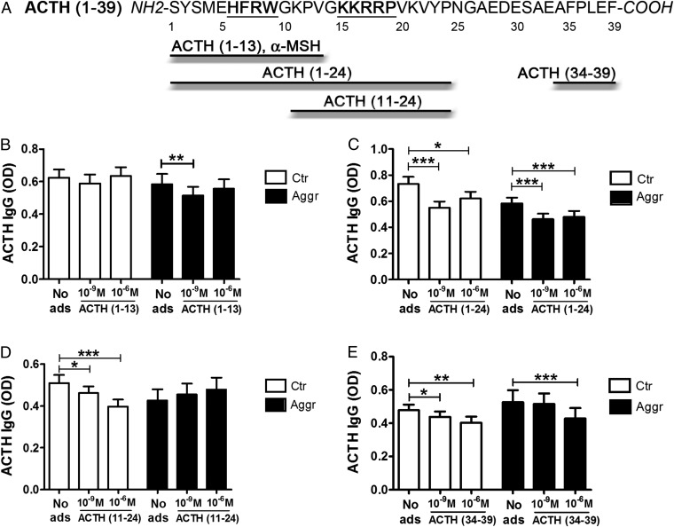
Fig. 3.
Epitope mapping of ACTH-reactive IgG in violent aggressors (Aggr) and healthy controls (Ctr). (A) Amino acid sequence of human ACTH (amino acids 1–39). The MC4R- and MC2R-binding sites HFRW and KKRRP, respectively, are underlined. Four different ACTH fragments used for plasma adsorption are shown. (B–E) Plasma levels in OD of IgG reactive with ACTH (amino acids 1–39) were measured before and after adsorption (ads) with 10−9 M and 10−6 M of each of the peptide fragments: (B) ACTH (amino acids 1–13); (C) ACTH (amino acids 1–24); (D) ACTH (amino acids 11–24); and (E) ACTH (amino acids 34–39). *P < 0.05, **P < 0.01, ***P < 0.001, paired t tests. Data are shown as mean ± SE; n = 20 control subjects and n = 16 violent aggressors.

