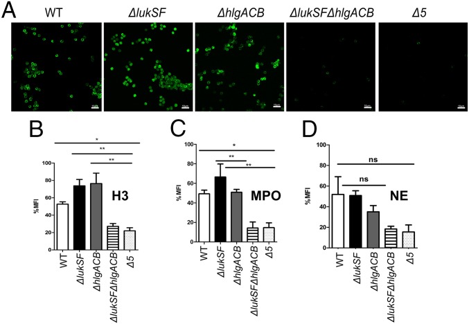
Fig. 3.
Biofilm-spent media containing PVL or HlgACB is sufficient for release of NETs. CLSM of neutrophils treated with biofilm-spent media of indicated strains, labeled with an anti-histone H3 (citrulline R2) antibody and Alexa Fluor 488 as a secondary antibody (green, A). Quantification of neutrophil staining with anti-citrullinated histone (B), anti-MPO (C), and anti-NE (D) antibodies after incubation with biofilm-spent media of indicated strains. ImageJ software 5.3 was used to calculate MFI per 100 cells for 10 fields from six independent experiments. Images were captured at a 600× total magnification and represent the majority population phenotype of six independent experiments performed in triplicate ± SEM (*P < 0.1, **P < 0.01, and ***P < 0.001, one-way ANOVA and Tukey’s post hoc analysis; ns, not significant). Neutrophils incubated with HBSS were used as a negative control. (Scale bars: 10 μm.)

