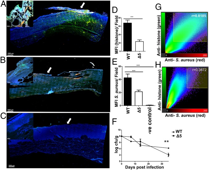
Fig. 5.
Leukocidins induce NETs and aid bacterial survival in vivo. Longitudinal sections taken across the wound bed of pigs infected with WT S. aureus USA300LAC (A) and the isogenic Δ5 strain lacking all five leukocidins (B) at day 7 post inoculation. Similar section taken from an uninfected control wound shown for comparison (C). Sections were stained with an anti-citrullinated histone antibody (green) and an anti-S. aureus antibody (red). Tissue cells were stained with DAPI and visualized at 100× total magnification. Images taken at 600× magnification from surface (white arrows) of corresponding wounds A–C (Inset). Mean fluorescence intensities of cells stained with antibodies against citrullinated histone and anti-S. aureus antibodies calculated for 10 independent fields taken across WT (D) and Δ5-infected (E) wounds. Counts of cfu per gram of tissue obtained by biopsy from WT and isogenic Δ5-infected animals (F). Pearson’s coefficients quantifying the degree of colocalization for anti-histone and anti-S. aureus antibodies for 10 fields of view, taken across WT (G) and Δ5-infected wounds (H). Results represent an average of two independent infections per strain performed in triplicate ± SEM (**P < 0.01 and ***P < 0.001, one-way ANOVA and Tukey’s post hoc analysis).

