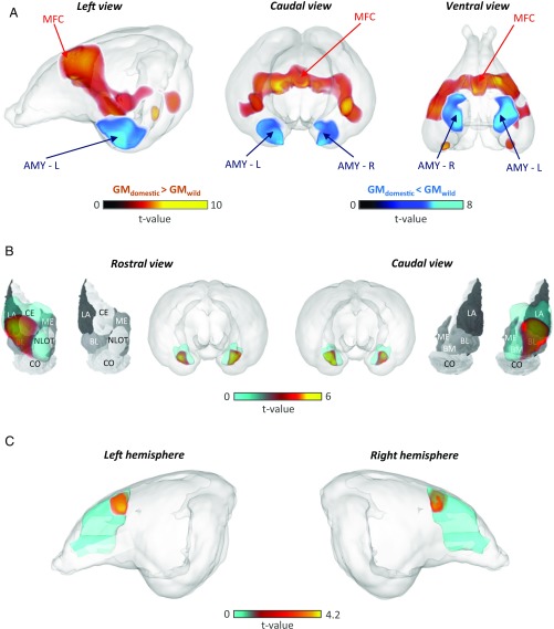
Fig. 3.
Specific changes in the size of the amygdala and prefrontal cortex between wild and domestic rabbits. (A) Amygdala (AMY-L and AMY-R, in blue) volume was smaller and medial prefrontal cortex (MFC, in red) volume was larger in domestic rabbits compared with wild rabbits. The two small areas with enhanced volume in domestic rabbits visible in the ventral view are not located entirely inside the cerebral region, but mainly intersect superficial vessel traces, and do not reflect meaningful GM changes. (B) The reduced amygdala volume in domestic rabbits compared with wild rabbits primarily concerns the basolateral (BL), lateral (LA), and central (CE) nuclei; these regions are denoted in red and superimposed on the rabbit nuclei map (11). (Modified from ref. 11.) (C) The medial frontal cortex ROI was enlarged bilaterally in domestic rabbits, with the maximum located dorsally as determined from VBM. The t value statistics in A–C were derived using the threshold free cluster enhancement method (33). L, left; R, right.

