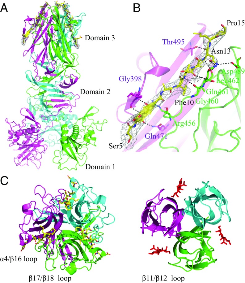
Fig. 3.
Crystal structure of the CofJ (1–24)–CofB complex. (A) Side view of the structure with the CofB trimer (cyan, magenta, and green) shown in ribbon representation and bound CofJ (1–24) peptides shown as yellow sticks. A 2mFo−DFc omit map contoured at 1.0 σ corresponding to the region of peptide-binding grooves is shown. (B) Peptide-binding interface of CofJ (1–24)–CofB complex. Close-up view of the Ser5–Pro15 fragment of one of the three CofJ (1–24) peptides. CofJ (1–24) peptide is depicted as yellow sticks. The residues of CofB participating in hydrogen-bonding interactions are shown in stick representation. Hydrogen bonds are shown as black dotted lines. (C) Structural comparison of domain 3 of CofB with CofJ (1–24) peptides (Left) and the H-type lectin domain of discoidin I with GalNAc molecules (Right, PDB code 2WN3). In the CofJ (1–24)–CofB structure, the individual monomers are colored in cyan, magenta, and green. In the discoidin I structure, the three bound GalNAc molecules are represented as sticks.

