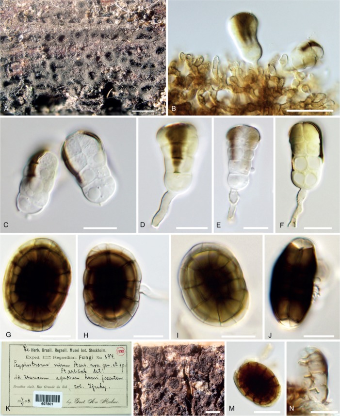
Fig. 11.

Hermatomyces tucumanensis (PMA 116083) A. Colonies on the natural substrate. B. Conidiogenous cells, young conidia and subicular hyphae. C. Young cylindrical conidia. D–F. Cylindrical conidia with still attached conidiogenous cell and conidiophore. G–J. Lenticular conidia. K. Envelope and content of the holotype of Scyphostroma mirum (BPI 697801). L. Colonies on the natural substrate. M. Lenticular conidium. N. Fragment of cylindrical conidium. Bar A = 500 μm, B–C = 20 μm, D–J = 10 μm, L = 500 μm, M–N = 10 μm.
