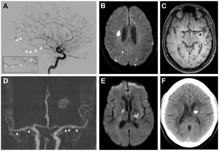
Figure 1.
Imaging of patients with PACNS. (A) A 44-year-old patient presenting with multifocal segmental narrowing of intracranial arteries on cerebral angiogram, (B) multiple DWI-lesions in different vascular territories, and (C) concentric enhancement of the M1-segment of the left middle cerebral artery on black blood MRI. (D) A 48-year-old patient with vessel beading on MRI-TOF-angiography, (E) bilateral infarctions of variable size (affecting different vascular territories and in various stages of healing), and (F) intracerebral haemorrhage.
DWI, diffusion-weighted imaging; MRI, magnetic resonance imaging; PACNS, primary angiitis of the central nervous system; TOF, time of flight.

