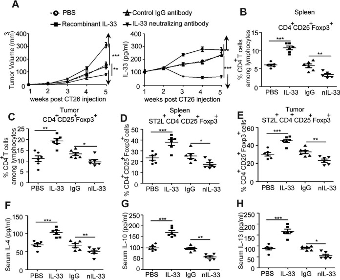
Figure 4.
IL-33 enhanced tumor size and antiinflammatory factor purified IL-33 and IL-33-neutralizing antibody were administered into tumor-bearing mice, and dynamic serum IL-33 levels or tumor volume were observed (1, 2, 3, 4, and 5 weeks after IL-33 injection via IV injection) (A). Single-cell suspensions of spleens or tumor tissues from control PBS group, IL-33 treatment group, control IgG-treated group, and IL-33-neutralizing antibody-treated group were prepared. Cells were stained with CD4-FITC, ST2L-APC, and CD25-PE-cyanine 7 and then intracellularly stained with PE-conjugated antibodies against Foxp3 for FACS analysis of CD4+CD25+Foxp3+ (Treg) (B, C) and ST2L+CD4+ CD25+Foxp3+ (ST2L+Treg) (D, E). The serum from above mice was purified for detecting the serum levels of Th2-related cytokine (IL-4, IL-10, and IL-13) by ELISA (F-H). ***P < .001, **P < .01, ANOVA/SNK. IL indicates interleukin; IV, intravenous; PBS, phosphate buffer serum; IgG, immunoglobulin G; Treg, regulatory T; PE, phycoerythrin; ELISA, enzyme-linked immunosorbent assay; FACS, fluorescence-activated cell sorting; ANOVA, analysis of variance; SNK, Student-Newman-Keuls.

