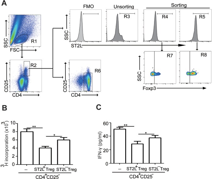
Figure 6.
ST2L+Tregs suppress CD4+CD25− T-cell proliferation and IFN-γ production single-cell suspensions of tumor tissue from tumor-bearing mice were prepared. Gating schemes for analysis of the percentages of ST2L+CD4+CD25+ cells (R5, gated from R1 and R2) or ST2L−CD4+CD25+ cells (R4, gated from R1 and R2) after purification by flow sorting (A) or spleen CD4+CD25− cells (R6, gated from R1) after purification by immunomagnetic sorting. Cells were stained with CD4-FITC, ST2L-APC, and CD25-PE-cyanine 7 and then intracellularly stained with PE-conjugated antibodies against Foxp3 for FACS analysis. Freshly sorted tumoral ST2L+CD4+CD25+ cells or ST2L−CD4+CD25+ cells were used in suppression assays, and the proliferation of spleen CD4+CD25− cells (B) or the IFN-γ secretion (C) was assessed with and without ST2L+CD4+CD25+ or ST2L−CD4+CD25+ cells at 1:2 ratio. The experiments were Performed twice, with similar results (**P < .01, Student t test). Treg indicates regulatory T; IFN-γ, interferon-γ; PE, phycoerythrin; FACS, fluorescence-activated cell sorting.

