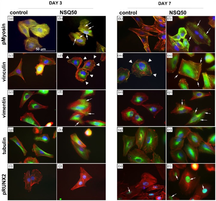
Figure 4.
Adhesion and cytoskeletal observations for SaOS2 cultured on planar control and NSQ 50 test surfaces after 3 and 7 days of culture. pMyosin at day 3 and day 7 showed enhanced stress fibre co-localisation for cells on NSQ 50 (b and l) compared to those of planar control (a and k, arrows). Considering focal adhesions, few were noted in cells on control at day 3 (c) compared to NSQ 50 at the same time point (d, arrow heads). By day 7, adhesions were noted in SaOS2 on control (m, arrowheads), but longer adhesions were observed in cells on NSQ 50 (n, arrows). On planar control, vimentin networks were observed around the nucleus of SaOS2 cultured on control at 3 (e) and 7 (o) days. On NSQ 50, however, the cells could be seen to have radiating vimentin networks extending to the cell peripheries at both days 3 (f) and 7 (p) of culture. At both time points and on both control and test materials, SaOS2 cells were seen to have well-organised microtubule networks (g, h, q and r). Considering pRUNX2 nuclear localisation at day 3, very little was noted on either the control (i) or NSQ 50 surface (j). However, at day 7, while nuclear localisation remained low in cells on control substrates (s), high levels of nuclear localisation was seen in cells on NSQ 50 (t, arrows).

