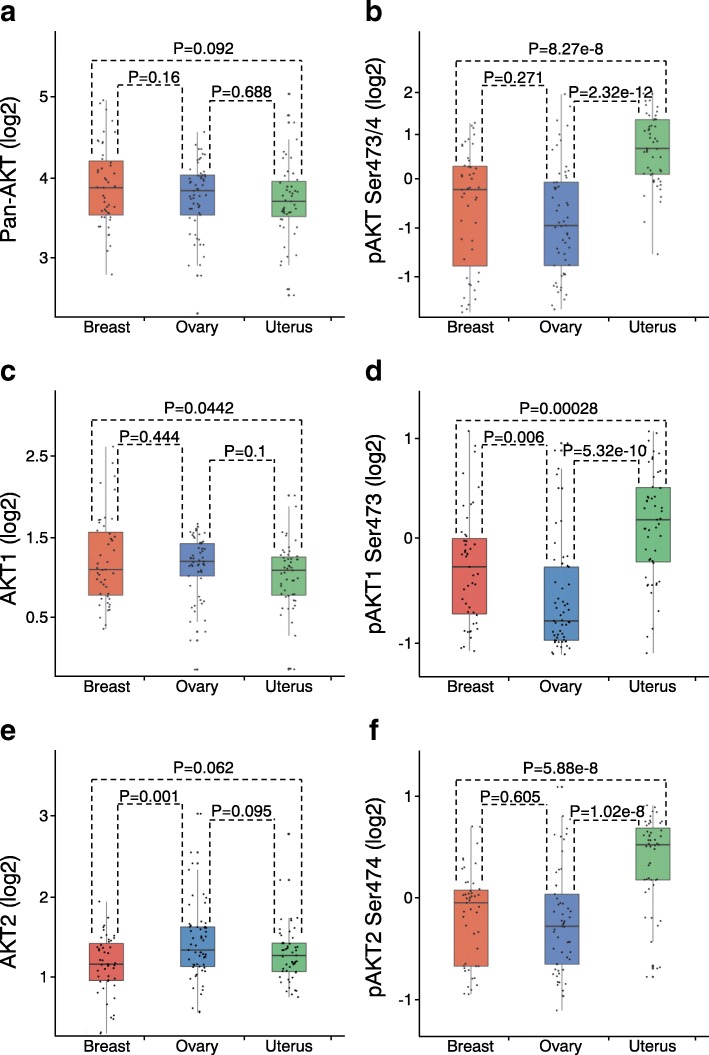
Fig. 3.
Differential AKT1 and AKT2 expression and activation across cancer lineages a, b Box plots of protein (a) or phospho-protein (b) levels of pan-AKT by cancer lineage. P values by t-test on log-transformed values. Box plots represent 5, 25, 50, 75, and 95%. c, d Box plots of protein (c) or phospho-protein (d) levels of AKT1. e, f Box plots of protein (e) or phospho-protein (f) levels of AKT2

