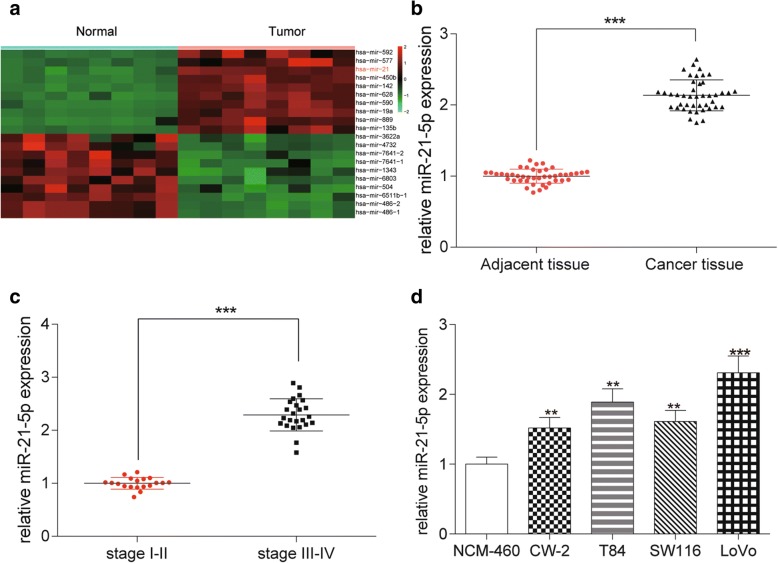
Fig. 1.
MiR-21-5p highly expressed in COAD tissues and cells. a The heat map showed that miR-21-5p was high expressed in COAD tissues compared with normal tissues. b The relative expression of miR-21-5p in cancer tissues was much higher than that in adjacent tissues detected by qRT-PCR. ***P < 0.001, compared with adjacent tissue. c The expression of miR-21-5p in stage III-IV was notably higher than that in stage I-II detected by qRT-PCR. ***P < 0.001, compared with stage I-II. d The expression of miR-21-5p in COAD cell lines (CW-2, T84, SW116, LoVo) was higher than that in normal colonic epithelial cell line (NCM-460) detected by qRT-PCR, which reached the highest level in LoVo cell line. **P < 0.01, ***P < 0.001, compared with NCM-460

