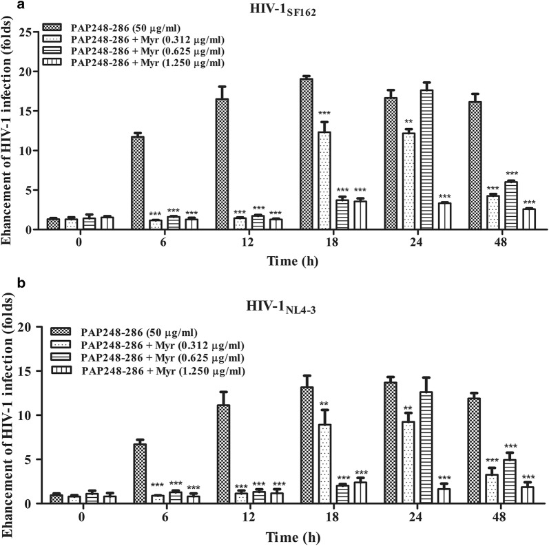
Fig. 2.
Amyloid fibril samples display loss of enhancement of HIV-1 infection in the presence of myricetin. Mixed SEVI fibril samples prepared as described above in the presence or absence of myricetin were diluted to a final concentration of 50 μg/ml (PAP248–286). The samples were then incubated with HIV-1SF162 (a) and HIV-1NL4-3 (b) infectious clones. The mixtures were added to prepared TZM-b1 cells. Luciferase activities were measured at 72 h post-infection. The values shown represent the mean ± SD (n = 3). One-way ANOVA with Dunnett’s post hoc multiple comparisons test was used to statistically analyze the differences between samples containing PAP248–286 alone and samples containing PAP248–286 and myricetin (*p < 0.05; **p < 0.01, ***p < 0.001)

